
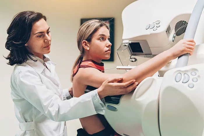
Breast cancer screening represents a critical component of women’s healthcare, with mammography remaining the gold standard for detection. Despite its proven effectiveness in identifying malignancies too small to palpate, traditional mammography presents several challenges, including discomfort during the compression process and significant rates of false positives.
The procedure requires flattening breast tissue between two plates while radiation captures images of internal structures. Many women find this experience physically uncomfortable and psychologically stressful, which contributes to screening avoidance, particularly among vulnerable populations. Current statistics indicate that approximately 85 percent of mammograms accurately detect cancers, though this percentage varies based on breast density and other individual factors.
Exploring Advanced Imaging Technologies for Improved Detection
Technological advancements are transforming breast cancer screening through various innovative approaches. Digital mammography utilizes computer-based imaging rather than traditional film, allowing technicians to enhance and manipulate images for better clarity. Magnetic resonance imaging employs powerful magnets and radio waves to generate detailed cross-sectional images of breast tissue, particularly useful for high-risk patients.
Ultrasound technology continues to evolve, with experimental three-dimensional imaging providing more comprehensive views of breast abnormalities. Emerging techniques like magnetic resonance elastography measure tissue stiffness, while microwave imaging spectroscopy detects variations in water content that may indicate malignant growths. These technologies aim to improve accuracy while reducing patient discomfort.
Investigating Minimally Invasive Cellular Diagnostic Procedures
Beyond imaging, significant research focuses on cellular-level detection methods. Ductal lavage represents a minimally invasive procedure where doctors insert a catheter into breast ducts to collect cells for microscopic analysis. This approach targets cancers originating in the milk ducts, potentially identifying malignancies before they become detectable through imaging.
Another experimental technique, ductoscopy, involves inserting a lighted scope into the ducts to visually examine the area for abnormalities. Researchers are also developing blood tests that analyze protein patterns to distinguish between malignant and benign conditions, with one study reporting 95 percent accuracy in detecting breast cancer, though these require further validation before clinical implementation.
Evaluating Experimental Electrical and Spectral Imaging Techniques
Several experimental screening methods utilize unique physical properties to detect breast abnormalities. Electrical impedance spectroscopy measures how breast tissue conducts low-frequency electrical current, creating images based on differences between normal and cancerous tissue conductivity.
Near-infrared spectroscopy exploits infrared light’s sensitivity to blood hemoglobin, generating images of vascular activity within breast tissue. Increased blood flow often indicates tumor growth, as malignancies require additional nutrients. Another approach, positron emission tomography, scans identify areas of high metabolic activity typical of rapidly dividing cancer cells. While these technologies show promise, most remain in clinical trials, requiring further validation before widespread adoption.
Addressing Screening Considerations for High-Risk Populations
Women with significant risk factors, including BRCA gene mutations, require specialized screening approaches. Genetic testing can identify mutations in BRCA1 and BRCA2 genes, which increase lifetime breast cancer risk to 50-85 percent. However, experts recommend thorough genetic counseling before testing due to potential psychological impacts and insurance discrimination concerns.
High-risk individuals may benefit from more frequent screening or alternative imaging like MRI, which offers superior sensitivity in dense breast tissue. Research continues to refine risk assessment tools and personalized screening schedules, balancing early detection benefits against potential overdiagnosis and unnecessary procedures.
Anticipating Future Directions in Breast Cancer Detection
The future of breast cancer screening likely involves multimodal approaches combining various technologies for optimal accuracy. Dartmouth College researchers are simultaneously investigating four screening techniques: near-infrared spectroscopy, microwave imaging spectroscopy, electrical impedance spectroscopy, and magnetic resonance elastography, with potential integration into a single device.
Blood-based biopsies represent another promising frontier, analyzing circulating tumor DNA or proteins to detect malignancies years before they become visible on imaging. While these advances offer exciting possibilities, researchers emphasize the need for rigorous validation through large-scale clinical trials and regulatory approval processes before clinical implementation.
Related topics:
