
Many women experience strange symptoms like heavy periods, pelvic pain, or a feeling of pressure in the lower belly. These signs can be caused by a common condition known as fibroids. Fibroids are non-cancerous growths in the uterus. They are very common, especially among women in their 30s and 40s. One of the best tools doctors use to detect fibroids is an ultrasound. But what do fibroids really look like on an ultrasound? Understanding this can help ease anxiety and give you more control over your health.
In this article, we’ll explain everything you need to know about fibroids and how they appear on ultrasound images. We’ll use clear and simple language so you can follow along easily, whether you’re preparing for an exam or just curious about your body. Let’s explore what those ultrasound pictures reveal and why they matter.
What Are Uterine Fibroids?
Uterine fibroids are benign (non-cancerous) tumors that grow in or around the uterus. They are made up of muscle and fibrous tissue. Fibroids can be as small as a pea or as large as a melon. Some women have just one, while others may have many.
Where Do Fibroids Grow?
Fibroids can grow in different areas of the uterus, such as:
- Intramural fibroids: Inside the muscular wall of the uterus.
- Subserosal fibroids: On the outer surface of the uterus.
- Submucosal fibroids: Just under the lining of the uterus.
- Pedunculated fibroids: Attached by a thin stem inside or outside the uterus.
Common Symptoms
Not all fibroids cause problems, but when they do, symptoms may include:
- Heavy or long menstrual periods
- Pelvic pain or pressure
- Frequent urination
- Backache or leg pain
- Difficulty getting pregnant
Why Ultrasound Is Used for Detecting Fibroids
Ultrasound is a safe and painless imaging test. It uses sound waves to create pictures of the inside of the body. It’s often the first step in diagnosing fibroids because it’s quick, affordable, and does not use radiation.
Types of Ultrasound
There are two main types of pelvic ultrasound used for fibroid detection:
- Transabdominal ultrasound: A probe is moved over the lower belly. This gives a wider view of the uterus.
- Transvaginal ultrasound: A slim probe is inserted into the vagina. This provides a closer and clearer view of the uterus and fibroids.
Benefits of Ultrasound
Ultrasound is often chosen because:
- It’s non-invasive
- It’s widely available
- It provides real-time imaging
- It’s safe during pregnancy
How Fibroids Appear on an Ultrasound
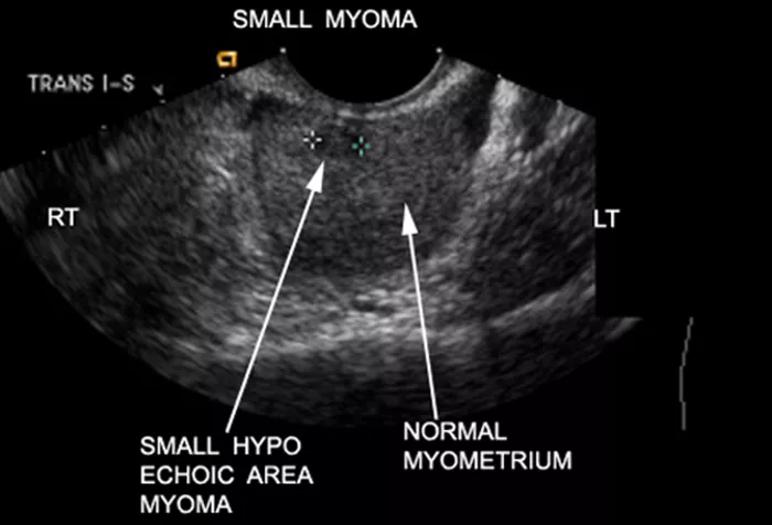
On ultrasound, fibroids have a distinct appearance. They usually show up as round or oval-shaped masses. The texture can vary depending on the type and age of the fibroid.
Fibroid Texture on Ultrasound
Fibroids often appear as:
- Hypoechoic areas: These are darker spots compared to the normal uterine tissue.
- Heterogeneous patterns: The texture may look uneven or mixed with different shades of gray.
- Shadowing: Fibroids may cause a shadow behind them due to the way they block sound waves.
Shapes and Borders
Most fibroids are well-defined and round or oval. Some may have irregular edges if they are degenerating (breaking down). Others may have small cyst-like spaces within them.
Size and Location
Ultrasound helps measure the size of the fibroid in centimeters. It also shows exactly where the fibroid is located, which helps guide treatment options.
Can Fibroids Be Missed on Ultrasound?
Sometimes small fibroids or those in tricky spots may be missed, especially with transabdominal scans. In such cases, doctors may suggest:
- Transvaginal ultrasound
- 3D ultrasound for better detail
- MRI if more information is needed
What Happens During the Ultrasound?
For a transabdominal scan, you’ll lie on your back with gel applied to your belly. The probe moves across the skin. For a transvaginal scan, the probe is gently inserted into the vagina. Both methods are usually painless and take less than 30 minutes.
What Your Doctor Looks For
During the scan, the doctor checks:
- The number of fibroids
- The size of each fibroid
- The shape and texture
- Whether they press on other organs
- Changes from previous scans
Ultrasound Report: What Do the Terms Mean?
Your ultrasound report may include technical terms. Here are some common ones:
- Hypoechoic mass: A dark area, often a fibroid.
- Heterogeneous texture: Uneven appearance inside the fibroid.
- Calcifications: Bright white spots from calcium build-up in older fibroids.
- Degeneration: Changes in the fibroid as it breaks down.
Do Fibroids Change Over Time?
Yes. Some fibroids grow, some shrink, and others stay the same. Pregnancy and menopause can affect their size. Regular ultrasounds can help track changes.
Can Ultrasound Help Decide on Treatment?
Yes. By showing how big the fibroids are and where they’re located, ultrasound helps doctors plan treatment. Treatment options may include:
- Watchful waiting (monitoring)
- Medications to shrink fibroids
- Minimally invasive procedures
- Surgery (myomectomy or hysterectomy)
When to See a Doctor
You should see a doctor if you have:
- Heavy bleeding
- Severe pelvic pain
- Rapidly growing belly
- Difficulty urinating or having bowel movements
- Trouble getting pregnant
Conclusion
Understanding how fibroids appear on an ultrasound can be a big step toward taking care of your health. These images are not just black-and-white shapes—they tell a story about what’s going on inside your body. Whether you’re having symptoms or just want peace of mind, ultrasound is a helpful and safe way to detect fibroids. Knowing what to expect during the scan and how to read the results makes the experience less stressful. Always talk to your doctor about your symptoms and ask questions about your scan results. The more you know, the more confident you can feel about your health and your options. Fibroids are common, but with the right care, they don’t have to control your life.
3 Common Questions About Fibroids and Ultrasound
1. Can fibroids be cancer?
No. Most fibroids are non-cancerous. Less than 1% turn into cancer, and this is very rare. Regular checkups help monitor changes.
2. How often should fibroids be checked with ultrasound?
If you have fibroids and no symptoms, you may only need a scan every 6 to 12 months. If you have symptoms, your doctor will decide how often to check.
3. Can ultrasound show if a fibroid is causing infertility?
Yes. An ultrasound can reveal if a fibroid is pressing on the fallopian tubes or changing the shape of the uterus, which can affect fertility.
READ MORE
