
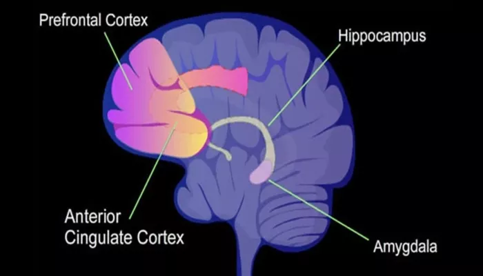
Researchers have identified a structural change in the brain that appears to track with relief from depressive symptoms following ketamine treatment. In patients with treatment-resistant depression, a reduction in the volume of a specific sub-region of the amygdala was linked to decreased feelings of sadness and unease. The findings were published in the Journal of Psychiatric Research.
Major depressive disorder affects millions worldwide. Standard treatments, such as selective serotonin reuptake inhibitors, help many patients, but roughly 30% fail to achieve sufficient relief even after multiple medications. These cases are classified as treatment-resistant depression.
Ketamine has emerged as a powerful alternative for these patients. Unlike traditional antidepressants, it acts through different neural pathways and can provide rapid relief for severe depression. However, the precise biological mechanisms behind its mood-improving effects remain unclear.
The amygdala, a small almond-shaped structure deep in the temporal lobes, plays a central role in processing emotions, especially fear and negative stimuli. In depression, this region often shows excessive activity, contributing to persistent negative moods. Functional imaging studies have shown that ketamine can reduce this overactivity, but the relationship between the amygdala’s physical volume and treatment response has been less well understood. Past research yielded inconsistent results, likely because the amygdala consists of multiple distinct subfields with different cellular structures and neural connections.
A team led by Kengo Yonezawa and Shinichiro Nakajima at Keio University School of Medicine in Tokyo hypothesized that examining these subfields individually could reveal links between structural changes and response to ketamine.
The researchers analyzed data from a double-blind, randomized, placebo-controlled trial—the gold standard in clinical research. Participants were adults aged 20 to 59 who had not responded to at least two antidepressants. Thirty-four patients were randomly assigned to receive either intravenous ketamine or a saline placebo twice a week for two weeks.
High-resolution MRI scans were conducted before treatment and five to six days after the final infusion. Using advanced software called FreeSurfer, the team segmented the amygdala into three subfields: the laterobasal nuclei, centromedial nuclei, and superficial nuclei. The laterobasal nuclei receive sensory inputs from the cortex, the centromedial nuclei send output signals to trigger behavioral responses, and the superficial nuclei connect to the olfactory cortex.
Depressive symptoms were measured using the Montgomery Åsberg Depression Rating Scale, including subdomains for dysphoria (sadness and pessimism), retardation (lethargy and lack of feeling), and vegetative symptoms (sleep and appetite changes).
The analysis revealed a specific correlation: in the ketamine group, a decrease in the volume of the right laterobasal nuclei was associated with reduced dysphoria. Patients who experienced more shrinkage in this region reported greater relief from sadness and unease. This effect was not observed in the placebo group, suggesting a drug-specific neurobiological change. No significant associations were found in other subfields or in the left amygdala.
These findings support the idea that overactivity in the amygdala contributes to depression. The laterobasal nuclei communicate with the prefrontal cortex, which regulates higher-order thinking and emotional control. Ketamine may restore this “top-down” regulation, with volume reduction reflecting a normalization of neural activity rather than a uniform shrinkage in all patients.
The study has limitations. The sample size was small, with 11 patients in the ketamine group and 15 in the placebo group. Participants remained on other antidepressants, which may have influenced results. The study was exploratory and did not include healthy controls, making it unclear whether volume changes reflect a return to normal size. MRI scans were taken shortly after treatment, so long-term effects are unknown.
Despite these limitations, the research offers a new perspective on ketamine’s action in depression. By linking structural changes in the right laterobasal nuclei to symptom improvement, it highlights a potential biological marker for treatment response. Understanding these markers could help develop personalized therapies and predict which patients will benefit from ketamine.
Related topic:
