
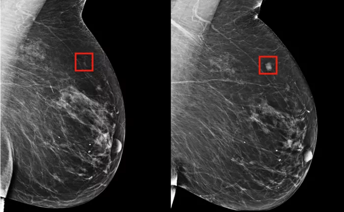
A newly developed artificial intelligence (AI) model has demonstrated remarkable accuracy in detecting tumor locations on breast MRI scans, outperforming existing benchmark models across multiple patient groups. The findings were published today in Radiology, the official journal of the Radiological Society of North America (RSNA).
Breast cancer screening traditionally relies on mammography, which, while widely accepted as the standard of care, has known limitations—particularly for women with dense breast tissue.
Dense breasts not only increase the risk of breast cancer but can also obscure tumors on mammograms, making detection more challenging. As a result, doctors often recommend breast MRI as an additional screening tool for women with dense breasts or those at elevated cancer risk.
According to Felipe Oviedo, Ph.D., senior research analyst at Microsoft’s AI for Good Lab and lead investigator of the study, breast MRI is more sensitive than mammography but comes with drawbacks. “MRI is more sensitive than mammography,” Dr. Oviedo explained, “but it’s also more expensive and has a higher false-positive rate, which can lead to unnecessary biopsies and patient anxiety.”
To improve the accuracy and efficiency of breast MRI screening, Dr. Oviedo’s team collaborated closely with radiologists at the University of Washington’s Department of Radiology to develop an explainable AI model focused on anomaly detection.
Unlike traditional models that attempt to classify images strictly as ‘cancer’ or ‘no cancer,’ anomaly detection models identify data points that deviate from what is considered normal, flagging these abnormalities for further review.
One significant challenge with previous AI models was their training data composition, often balanced evenly between cancer and non-cancer cases. “Such a 50-50 split is unrealistic for screening populations where cancer prevalence is very low, typically around 2% or less,” Dr. Oviedo said. Additionally, earlier models lacked interpretability—making it difficult for clinicians to understand or trust their outputs, which is crucial for clinical adoption.
To address these challenges, the research team trained their AI model on nearly 10,000 consecutive contrast-enhanced breast MRI exams conducted at the University of Washington from 2005 to 2022. The patient cohort was predominantly white (over 80%), with 42.9% having heterogeneously dense breasts and 11.6% categorized as having extremely dense breast tissue.
The model learned a comprehensive representation of benign breast tissue, enabling it to better recognize abnormal malignancies even when such cases were scarce in the training data. Dr. Oviedo emphasized, “Malignancies can appear in many different ways, and because they are relatively rare in these datasets, our anomaly detection approach is a promising solution for real-world screening.”
This AI-assisted breast MRI technique holds potential to uncover cancers that may be missed by human radiologists alone, especially in women with dense breast tissue where mammography struggles. It could also reduce false positives and improve the overall effectiveness of breast cancer screening.
While further validation and clinical trials are needed before widespread implementation, this study marks an important step forward in integrating AI into breast cancer diagnostics, potentially enhancing early detection and patient outcomes.
