
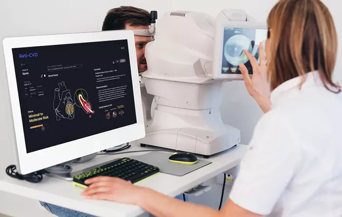
A routine visit to the optometrist often involves placing your chin and forehead against a bioimaging device known as optical coherence tomography (OCT).
Widely used in eye clinics worldwide, OCT employs light waves to capture high-resolution, cross-sectional images of the retina without invasive procedures—critical for diagnosing and monitoring a range of eye conditions.
Traditional OCT devices rely on mechanical components, such as spinning mirrors, which can increase the risk of device failure and limit long-term reliability.
Recognizing these challenges, researchers at the University of Colorado Boulder have developed a novel bioimaging device that operates without mechanical parts and requires significantly less electrical power. Their innovation could enhance detection of both eye and heart conditions.
Published in Optics Express, the study details a device that leverages electrowetting—a process that alters the surface shape of a liquid to perform optical functions. By eliminating scanning mirrors, the device reduces energy consumption while minimizing mechanical wear and extending operational lifespan.
“The benefits of non-mechanical scanning are substantial,” said the team’s lead engineer, Gilinsky. “Without moving parts, the device is less prone to mechanical failure and more durable overall.” He emphasized that OCT systems must be compact, lightweight, and safe for human use.
The research team included Juliet Gopinath, professor of electrical engineering; Shu-Wei Huang, associate professor of electrical engineering; Victor Bright, professor of mechanical engineering; PhD graduates Jan Bartos and Eduardo Miscles; and PhD student Jonathan Musgrave.
“Our work opens the door to earlier detection of health conditions, ultimately improving patient outcomes,” said Gopinath.
Testing with Zebrafish
To validate the device’s biomedical imaging capabilities, the researchers turned to zebrafish, whose eye structures closely resemble those of humans. Focusing on the cornea, iris, and retina, the team achieved axial resolution of 10 microns and lateral resolution of approximately 5 microns—smaller than the width of a human hair.
“The exciting outcome was our ability to clearly delineate the cornea and iris, meeting our resolution goals,” Gilinsky noted.
Accurate imaging at this scale could revolutionize early diagnosis of eye diseases such as age-related macular degeneration and glaucoma. Beyond ophthalmology, the technology may also assist in mapping human coronary features, providing valuable insights for heart disease—the leading cause of death in the United States.
With their expertise in microscopy systems, the researchers envision future applications in minimally invasive endoscopes. “There’s a growing demand for endoscopes that are small, flexible, and comfortable for patients. Our non-mechanical optical components allow us to maintain high performance in a compact form factor, advancing OCT technology,” Gilinsky explained.
READ MORE
FDA Approves New Eye Drops to Help Adults See Up Close Better
