
Fracture healing typically takes months, with doctors currently relying on periodic 2D X-ray images to monitor bone regeneration. Patients require repeated scans every few weeks or months until complete healing is evident.
Particularly concerning are tibia fractures, which experience delayed or stalled healing in up to 25% of cases, with underlying conditions like age or diabetes further complicating recovery. Traditional methods struggle with early detection of healing abnormalities, potentially leading to prolonged pain and extended medical interventions.
Technological Innovation and Research Vision
A five-year, $3 million project led by University of Delaware mechanical engineer Michael Hast aims to develop radiation-free imaging technology for early identification of bone healing issues.
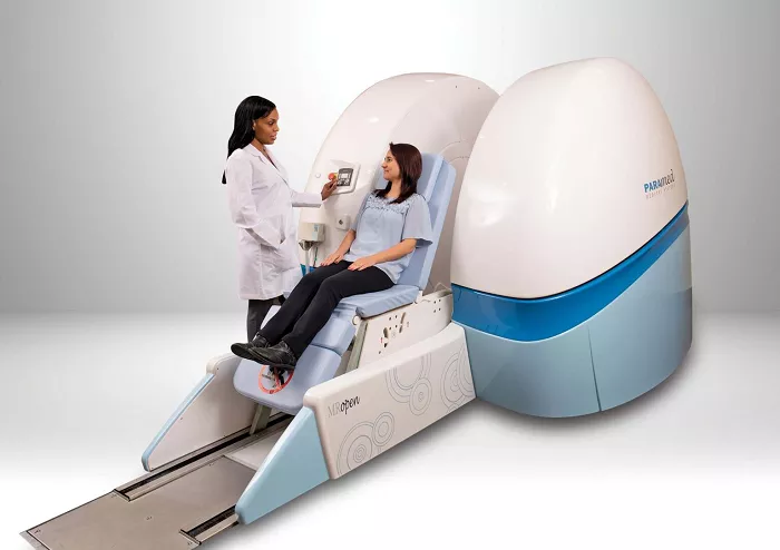
Supported by an NIH R01 grant, the research pioneers a novel approach combining ultrashort echo time MRI with 3D computational modeling to assess healing bone strength without ionizing radiation exposure. This initiative represents a paradigm shift from reactive monitoring to proactive intervention in fracture management.
Technical Methodology and Interdisciplinary Collaboration
The research team is developing a groundbreaking method that maps MRI data to three-dimensional voxels to create computational models simulating real bone mechanical properties. Professor Hast collaborates with modeling expert Hannah Dailey from Lehigh University and University of Pennsylvania surgeons in this interdisciplinary effort.
The team first validated their approach using sheep bone healing models, comparing MRI-based predictions against laboratory measurements of mechanical strength.
Clinical Validation Protocol
The project plans to enroll approximately 50 tibial fracture patients at the University of Pennsylvania for a year-long longitudinal study. Researchers will track participants’ recovery progress while comparing MRI model predictions against actual healing outcomes.
This comprehensive validation design will assess the technology’s effectiveness in predicting human fracture repair progression and identifying early warning signs of compromised healing.
Clinical Applications and Immediate Benefits
The technology promises to help clinicians quickly evaluate new bone development and determine whether it can withstand stresses from daily activities. “Better predictive tools can give clinicians and patients greater confidence that healed bone can withstand physical activity without refracture risk,” Hast noted.
“We aim to detect early warning signs of poor healing so providers can adjust rehabilitation protocols sooner, getting patients back on track faster.” Recent evidence suggests early mobilization benefits leg fracture recovery, and this technology could provide scientific justification for such approaches.
Long-term Impact and Field Transformation
Successful implementation could transform fracture management from passive observation to active intervention. The radiation-free monitoring technology, while initially focused on tibial fractures, holds potential for adaptation to other fracture types.
Project outcomes may significantly reduce healthcare costs, improve patient quality of life, and usher orthopedics into an era of personalized rehabilitation medicine through advanced predictive modeling and early intervention capabilities.
Related topics:
