
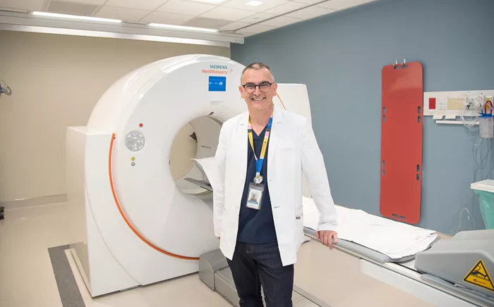
A large multicenter study led by the London Health Sciences Centre Research Institute (LHSCRI), the Lawson Health Research Institute, and the University Health Network (UHN) has identified a powerful new imaging technique that significantly improves the detection of recurrent prostate cancer and is linked to better survival outcomes. The study, conducted over seven years, has been published in The Journal of Nuclear Medicine.
The imaging approach, known as prostate-specific membrane antigen (PSMA) positron emission tomography (PET) scanning, allows clinicians to locate prostate cancer recurrences much earlier and more precisely than conventional imaging methods.
During the procedure, a radioactive tracer designed to bind specifically to a protein expressed on prostate cancer cells is injected into the patient’s bloodstream prior to scanning. This enables clinicians to visualize even small clusters of cancer cells that standard scans often miss.
“This new technique gives physicians the information they need to select the most effective treatment,” says Dr. Glenn Bauman, Scientist at LHSCRI and Radiation Oncologist at London Health Sciences Centre. “When blood tests suggest a recurrence but standard imaging fails to identify where, doctors are often forced to use broader, less targeted therapies. PSMA PET scans let us pinpoint the cancer and deliver focused treatment instead.”
The study reported an impressive 70% overall detection rate, compared with historical detection rates of only 10–20% using traditional bone or CT scans. Furthermore, the results led to a change in treatment management for approximately half of the participants. Among men with cancer detected by PSMA PET, nearly 90% had their treatment plans adjusted following the scan. Importantly, patients whose care was modified based on PSMA PET results demonstrated significantly improved overall survival compared to those assessed with standard imaging alone.
“We’re encouraged by how this imaging approach is already reshaping cancer care,” says Dr. Ur Metser, Division Head of Molecular Imaging at UHN and Clinician Scientist at the Princess Margaret Cancer Centre. “Our data show that PSMA PET scanning leads to more personalized treatment decisions — and those individualized choices are associated with longer survival. It’s a meaningful step forward for both patients and their care teams.”
Dr. Bauman and his colleagues were the first in Canada to use PSMA PET imaging in a patient back in 2016. Since then, the ongoing study has involved thousands of men across six hospitals in Ontario, supported by funding from Ontario Health – Cancer Care Ontario. Based on the strength of these findings and complementary research, PSMA PET scans have now been approved and publicly funded as a standard-of-care diagnostic test for men with advanced prostate cancer in Ontario.
Related topics
