
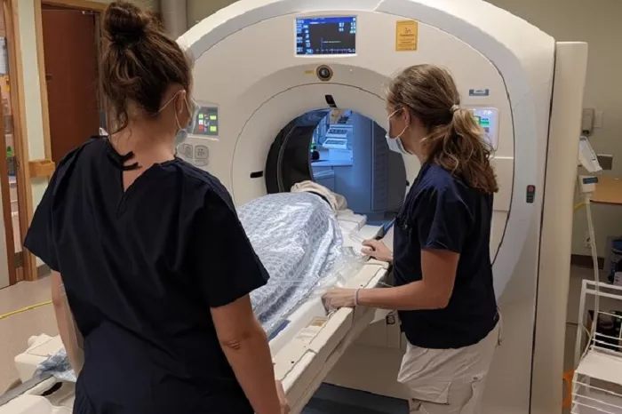
A new study published online in Radiologyon November 11 demonstrates that artificial intelligence (AI) can repurpose routine chest, abdominal, and spine CT scans—originally performed for conditions like kidney stones or lung abnormalities—to detect signs of bone loss and osteoporosis.
Developed by radiologists at NYU Langone Health in collaboration with Visage Imaging, this AI tool enables opportunistic screening for osteoporosis using existing medical imaging data, potentially identifying at-risk patients who would otherwise remain undiagnosed.
Study Design and Methodology
The research team analyzed 538,946 CT scans from 283,949 NYU Langone patients, utilizing 43 different scanner models and all standard testing protocols to ensure broad applicability. The AI tool measured bone mineral density at specific thresholds for each major lumbar and thoracic vertebra, with calculations adjusted for age, sex, race, and ethnicity. Radiologists subsequently validated the accuracy of the AI-generated findings against clinical standards.
Key Findings and Demographic Trends
The analysis revealed significant patterns in bone density across diverse patient populations:
Age and Sex Dynamics: Women under the age of 50 had higher bone density than men of the same age, but this advantage reversed after menopause, with men over 50 exhibiting higher bone density than older women
Racial and Ethnic Variations: Black individuals had the highest bone density, followed by Asian and white populations
Clinical Utility: AI-identified bone density thresholds effectively flagged weakened bones, enabling early intervention opportunities
Clinical Implications and Public Health Impact
“By leveraging the vast amount of imaging data we already collect, we can address the underdiagnosis of osteoporosis and help people build stronger, healthier bones,” said co-investigator Dr. Soterios Gyftopoulos, professor of radiology and orthopedics at NYU Grossman School of Medicine.
Previous research by Gyftopoulos estimates that opportunistic screening could double the number of patients tested annually for osteoporosis, potentially saving over $2.5 billion in Medicare costs each year. With osteoporosis affecting more than 10 million Americans—mostly women over 50—and an additional 40 million showing early signs of low bone mass, this approach addresses a critical gap in preventive care.
Implementation and Future Directions
The AI tool will soon be deployed in a clinical trial at NYU Langone hospitals, using CT scans originally performed for other purposes to screen patients with unknown bone density status.
Senior investigator Dr. Miriam Bredella, Bernard and Irene Schwartz Professor of Radiology at NYU Grossman School of Medicine, emphasized that widespread adoption could change the disease trajectory for millions who remain undiagnosed until experiencing fractures—some of which, like hip fractures, can be life-threatening.
Broader Applications and Research Expansion
The NYU Langone team plans to develop similar AI-based programs using existing scan data to diagnose other conditions, including cardiovascular disease and muscle mass loss. Early research already indicates that opportunistic screening of abdominal CT scans can reveal cardiovascular risks, demonstrating the broad potential of repurposing routine imaging for preventive health initiatives.
Collaboration and Funding
The study was supported by NYU Langone Health, with collaborative contributions from Visage Imaging in Berlin, whose AI tools helped develop the osteoporosis screening program. The research represents a growing trend toward leveraging existing medical data and AI technologies to enhance disease detection and reduce healthcare costs.
Related topics:
