
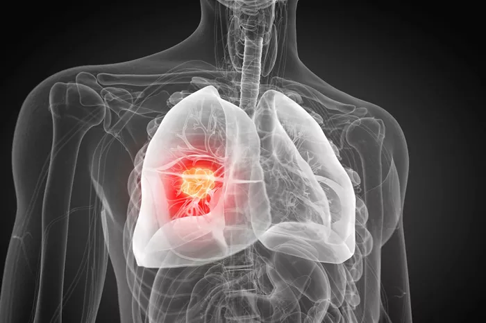
In the fight against lung cancer, precision is paramount. Radiation therapy relies on accurately identifying and mapping tumors to deliver high-dose radiation that destroys cancer cells while protecting healthy tissue.
Traditionally, this crucial task—known as tumor segmentation—has been performed manually by oncologists. This process is time-consuming, varies between doctors, and can sometimes miss critical tumor areas, potentially affecting patient outcomes.
A pioneering AI tool named iSeg, developed by Northwestern Medicine scientists, promises to change this landscape. According to a recent large-scale study published in npj Precision Oncology, iSeg matches expert physicians in outlining lung tumors on CT scans and can even detect tumor regions that some doctors overlook.
What Makes iSeg Different?
Unlike earlier AI models that analyze static images, iSeg is the first 3D deep learning system capable of segmenting tumors as they move with each breath. This dynamic approach is vital because lung tumors shift position during respiration, and accounting for this movement improves the accuracy of radiation targeting. Given that half of all cancer patients in the U.S. receive radiation therapy, this advancement could have widespread impact.
Proven Accuracy Across Multiple Clinics
The development team trained iSeg using CT scans and tumor outlines from hundreds of lung cancer patients treated at nine different clinics within the Northwestern Medicine and Cleveland Clinic systems. This extensive dataset surpasses the limited, single-hospital data used in many previous studies, enhancing the AI’s robustness.
When tested on new patient scans, iSeg consistently matched expert tumor contours and identified additional suspicious areas that some physicians missed. Importantly, these missed regions were linked to poorer patient outcomes if left untreated, highlighting iSeg’s potential to improve survival rates by catching high-risk tumor zones early.
Benefits for Patients and Healthcare Providers
Accurate tumor targeting is essential for effective radiation therapy. Even small errors can reduce tumor control or cause unnecessary damage to healthy tissue. By automating and standardizing tumor segmentation, iSeg can:
Reduce delays in treatment planning
Ensure consistent quality of care across hospitals
Identify hidden tumor areas that may be overlooked
Ultimately improve patient outcomes and safety
Next Steps: Clinical Integration and Expansion
The research team is currently testing iSeg in real-time clinical settings to compare its performance directly with physicians. They are also working to incorporate user feedback and expand the technology to other cancers, including liver, brain, and prostate tumors. Future versions aim to support additional imaging methods like MRI and PET scans.
Troy Teo, radiation oncology instructor and co-author, envisions iSeg as a foundational tool that could standardize tumor targeting in radiation oncology nationwide. He predicts clinical deployment within just a few years, potentially transforming cancer care by making precise, personalized treatment accessible to more patients.
Related topics:
